

EyeCare Specialties of Illinois
Our board-certified eye doctors provide the highest quality of care performing everything from routine eye exams to diagnosing, treating, and managing eye diseases.
Find an Eye Doctor Near You
About EyeCare Specialties of Illinois
At EyeCare Specialties of Illinois, our top priority is customer care. Our family of practices has been caring for local communities for decades. Our board-certified doctors provide the highest quality of care, performing everything from routine eye exams to diagnosing, treating, and managing eye diseases.
Our knowledgeable opticians will help you choose the best frames to fit you and your lifestyle. Each practice in the EyeCare Specialties network carries a range of designer eyewear collections for every family member and every budget. Our knowledgeable, friendly staff is ready to serve you at each of our practices. Come in, call, or schedule your eye exam online.

Find an Eye Doctor Near You
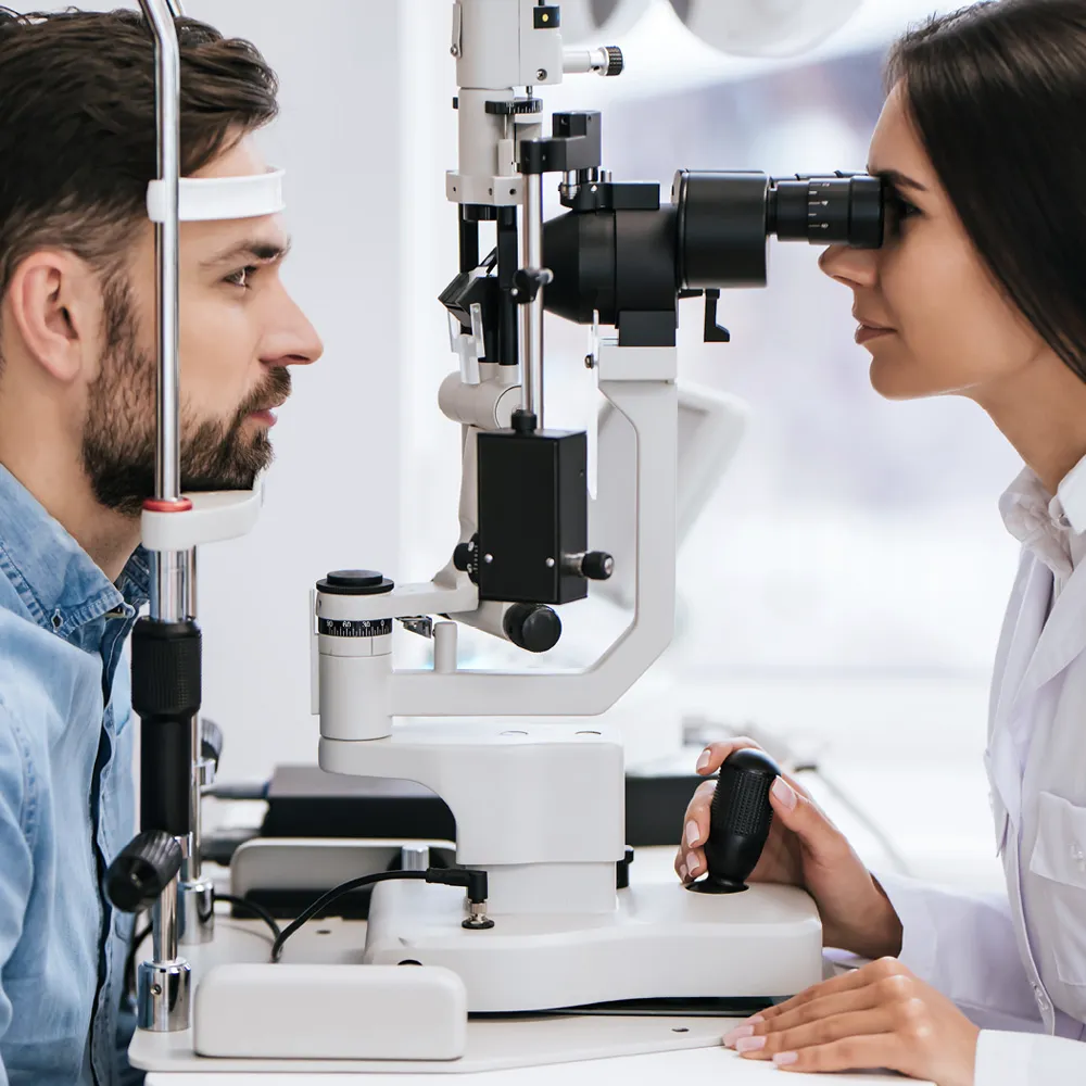
At EyeCare Specialties of Illinois we believe in using the latest technologies to aid us in doing everything comprehensively—right down to your basic eye exam.